Secondary Xylem and Tyloses in Stems

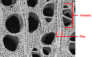
In the scanning electron micrograph image at left, three secondary xylem vessels are positioned side-by-side. Lateral movement of water and minerals diffuse through pits on their side walls.
A ray composed of ray parenchyma cells is also shown.

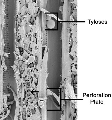
Tyloses are common in Vitis. A tylose is formed by a parenchyma cell that has grown through the pit of a vessel member.
Below the tyloses in the vessel at right is a simple perforation plate. This plate indicates where two vessel members have joined together to form a continuous conducting tube.
The arrow depicts amyloplasts enclosed in a ray parenchyma cell. Amyloplasts are starch storing plastids.
Note the bordered scalariform pits on the walls of the vessel at left.