Photos Provided by Dr. Geoff Burrows
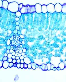
The leaf TS is about 200 um thick and shows:
relatively large upper epidermal cells with thin cuticle and no stomata
single layer of densely packed palisade mesophyll
relatively densely packed spongy mesophyll
vascular bundle with bundle sheath and support tissues
lower epidermis with two pairs of guards cells and associated stomata plus substomatal cavities .
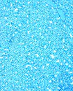
The palisade paradermal shows the relatively dense packing of the cells but also shows there is a well developed system of air spaces for gas exchange.
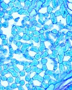
The spongy mesophyll paradermal shows a greater amount of intercellular airspace. The palisade and spongy mesophyll images are good to present side by side.
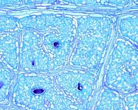
The paradermal of the veins shows the reticulate nature of the vascular system, the ‘blind' endings of some veins and that all leaf cells are in relatively close proximity to xylem/phloem.
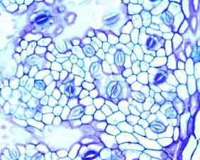
The lower epidermis paradermal gives another view of the stomatal complexes.
Dr Geoff Burrows ( mailto:GBurrows@csu.edu.au )
Senior Lecturer
Strand Advisor BSc (Hort)
School Ag & Vet Sci
Charles Sturt University
Locked Bag 588, Wagga Wagga 2678
ph 02 69 332 654 fax 02 69 332 812