|
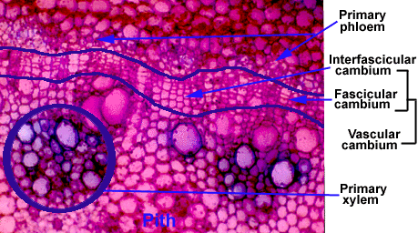
This cross section was taken from a stem younger than the ones you have just seen. Here, secondary growth is just beginning. The vascular cambium is visible, but no significant amount of secondary tissue can be seen. The vascular cambium is recognizable in cross section because the cambial cells always have a flattened appearance. To see for yourself, compare the cells labelled as vascular cambium with the cells in the pith. |

|
|
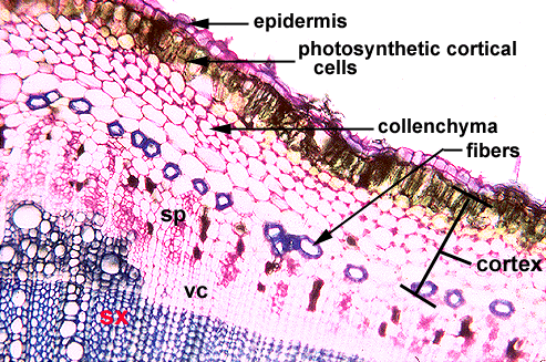
The following photograph is a cross section of stem with considerable secondary growth. The secondary xylem and phloem have been labelled. Note how the cortex is still intact. You can even see cells that are still photosynthetic at the surface of the stem, and some collenchyma tissue is present. Note also the fiber cells that are located at the boundary between the secondary phloem and the cortex. [sp=secondary phloem; vc=vascular cambium; sx=secondary xylem] |

|
|
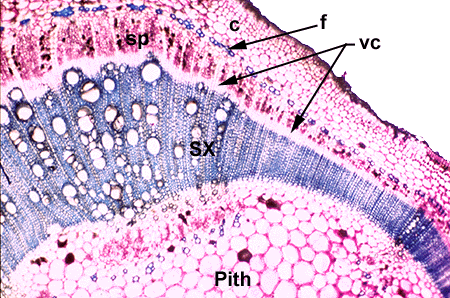
This final cross section is at a lower magnification and includes the pith. You can see again the uneven secondary xylem growth. In this stem the epidermis and outer cortex appear to be deteriorating. This is common in stems that are undergoing secondary growth. [sx=secondary xylem; vc=vascular cambium; f=fiber; c=cortex] |

To go back to the stem secondary growth main page click here. To look at tangential longitudinal sections of secondary xylem (wood) click here. |