| Secondary growth occurs in many roots and usually results in the thickening of the root diameter by the addition ofvascular tissue. | |
|
Initiation of secondary growth occurs when cells in the residual procambium and parts of the pericyle begin to make periclinal divisions. Only the pericyle cells opposite the xylem points start to make periclinal divisions. The inner layer of cells becomes the vascular cambium. The outer layer is retained as pericycle. The vascular cambium is continuous around the primary xylem. |
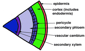
| The vascular cambium continues to divide periclinally. The daughter cells that result from these divisions differentiate into secondary xylem cells if they divide off towards the inside of the root or secondary phloem cells if they divide towards the outer surface of the root. After many cell divisions and cell differentiation, a root exhibiting secondary growth might look like the one depicted in the diagram to the right. |
|
| Some roots also form an outer protective layer called the periderm which originates from the pericycle and replaces the epidermis. | |
|
The pericycle resumes its meristematic character and begins to divide periclinally again. At this point it is called the phellogen or the cork cambium. This diagram, which is a slice of the previous drawing, shows how the pericycle is dividing. |
|
The cork cambium forms phellum cells (cork cells) towards the outside of the plant. These cells are dead at maturity. They are suberized which makes the cells impermeable to water. Phellum cells in cross section appear in neat, ordered files. The cork cambium also produces the phelloderm, a tissue consisting of cells that are living at maturity. The location of the phelloderm can be seen in the diagram to the left. |
| Not all plants have secondary growth in their roots, and many do not conform to the developmental pattern that was described above. | |
| Tomato roots do exhibit secondary growth, however they do not form a clear periderm. The photographs below show what secondary growth looks like in the tomato roots. The vascular cambium can be difficult to spot at a low magnification, but at a higher magnification, one can see it more clearly by noticing the periclinal divisions in this layer which result in cells that have a "squashed" appearance. | |
|
|
|