|
|
|
|
|
For some general information about secondary growth in roots, visit the tomato page. 
Secondary growth in roots develops approximately 12 centimeters behind the root apex. Several anatomical changes occur during secondary growth including the addition of vascular tissue and the loss of the cortex and the epidermis which are replaced by a multi-layered periderm.
|
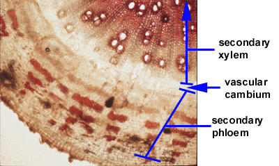
Secondary tissues have a distinct appearance. In the following photograph of a cotton root cross section (stained with phloroglucinol, a lignin stain), you can see that the secondary xylem is composed of large lignified cells (xylem vessels) which stain red. The secondary phloem which is situated outside the xylem is made of smaller cells, mostly sieve tube members, companion cells, phloem fibers, and parenchyma. In between the secondary xylem and the secondary phloem is the secondary meristem, the vascular cambium, which produces those two tissues. The vascular cambium divides in the periclinal plane (parallel to the surface of the root) to produce secondary vascular tissues and divides anticlinally (perpendicular to the root's surface) to increase its own circumference. The result of these divisions is an increased girth of the root.

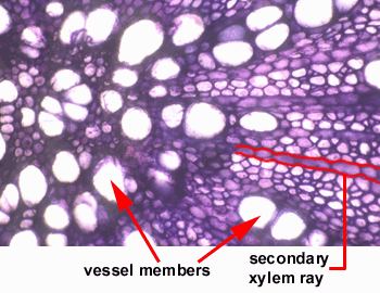
The photograph below is of a cross section through a cotton root with secondary growth. What you see is secondary xylem that has been stained with toluidine blue. A secondary xylem ray has been labeled. Rays are made up of parenchyma cells and are useful in lateral transport within the root. Can you find any other rays in this photograph? Two large xylem vessel members have been identified. Not all vessels are that large. Many of the smaller cells in this section are xylem vessel members too. 
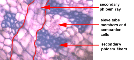
The next photograph is a close-up cross sectional view of secondary phloem. The most prominent cells in this section are the phloem fibers which stain blue with the toluidine blue stain. The cells surrounding the patches of fibers are sieve tube members and companion cells. A phloem ray, made of parenchyma cells has been identified as well. A pattern of alternating patches of fibers and conductive phloem cells was observed in cotton. 
|
|
Introduction | Flowers&Fruit | Roots | Stems | Leaves Section of Plant Biology Division of Biological Sciences UNIVERSITY OF CALIFORNIA, DAVIS |