| Roots can have open or closed organization at the root tip. How a root tip is organized can best be seen with a median longitudinal section. | |
|
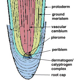
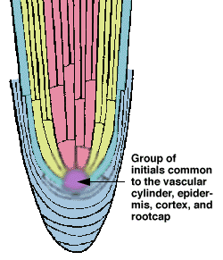
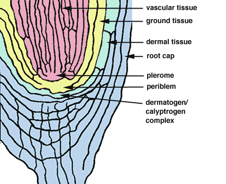
Closed organization means that the files of cells that arise from the root tip can be traced back to meristematic layers, or histogens. For example, the vascular tissue in this diagram can be clearly traced back to a single layer of cells. This histogen is called the plerome. The ground tissue in this drawing originates from a layer called the periblem, and the epidermis and the root cap both share a histogen layer which is called the calyptrogen/dermatogen complex. |
| In open organization, the cell files cannot be traced back to a single, distinguishable layer. |
|
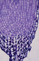
| Tomato roots have closed apical organization. In the photograph below, note how the cells form layers as they approach the root tip. The line drawing, representing the photograph, indicates cell rows and also demonstrates how the tomato root cell files converge into a single layer at the tip of the root. | |
|
|
| Line drawing courtesy of Dr. David | |
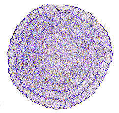
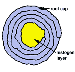
| A cross section through a tomato root tip, reveals an interesting feature that can also be found in many other plants. The root cap cells in the tomato appear to be organized into a spiral. Note that the continuous spiral of cells terminates at a region where the cells seem to have no clear pattern. This area (represented by yellow in the line drawing) is a histogen layer. The spiral is the result of a specific developmental cell division pattern. | |
|
|
| Special thanks to Sue Nichol for the the root cap sections. | |