|
We shall now examine some cell types and discuss their functions.
|
|
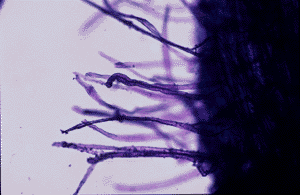
| These are root hairs from the tomato root. They are single cell extensions in the root epidermis that function in the absorption of water and nutrients. Root hairs increase the surface area over which the plant can absorb water and nutrients. |
|
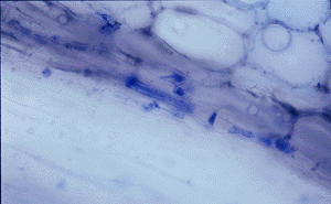
| The cell with blue plugs on both ends is a sieve tube member(STM). A strand of STMs connected end to end forms a sieve tube which makes up part of the phloem tissue of the plant. Sieve tubes function in the transport of carbohydrates, amino acids, and proteins, among other substances that are crucial to the plant's survival. |
|
| Xylem tissue functions mainly in the transport of water from the roots to the shoot of the plant. This tissue can be made up of several cell types. However, the xylem vessel members compose most of the xylem tissue. Two types of xylem vessel members can be described. | |

|
The photograph to the left shows a protoxylem vessel member taken from a tomato root. The secondary wall around this cell is a spiral. The protoxylem tissue matures before metaxylem (see photograph below) tissue does. Protoxylem allows for more flexibility than the metaxylem which is an important feature in the growing parts of the plant. |
|
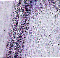
The metaxylem vessel member is on the left side in this photograph. Notice how there is much more secondary wall material in the metaxylem vessel member than there is in the protoxylem vessel member next to it. This makes the cell more rigid. Also notice the pits in the cell walls. These pits allow for the lateral movement of water within the plant. |
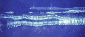
| This photograph of a longitudinal section through a root shows a protoxylem vessel member with a unique double helical secondary wall thickening. |
|
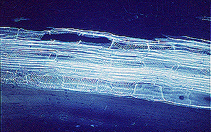
| This photograph shows how the vessel members connect to form a continous vessel. There is no cell wall material at the ends of the vessels (tomatoes have a simple perforation plate) so, there is no resistance to water at the junction of two vessels. |
|
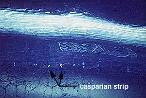
| Another specialized cell in the root is the endodermal cell. The endodermis forms a cylinder around the vascular cylinder in the roots. The endodermal cells have a suberin layer called the casparian strip embedded into their cell walls. This suberin can be stained using a special dye and appears when viewed under fluorescent light. The casparian strip appears as light spots in the endodermis in this longitudinal section through the root, but the suberin is laid down in a continuous layer that cannot be seen in such a section. This photograph also provides a nice example of a metaxylem vessel member. |
|