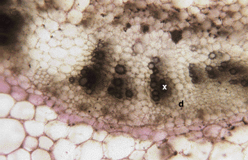
 cross section through petiole (x-xylem, d-phloem) |
| This shows a closer view of the xylem and phloem inside the petiole. Notice that the xylem vessels have a very thick secondary wall. This offers the vessel the support it needs to withstand the tension created as the water is drawn up from the roots. |
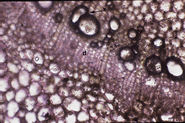
 cross section through petiole (x-xylem, d-phloem) |
| This cross section shows us an even closer view of the vascular tissue inside petioles. |
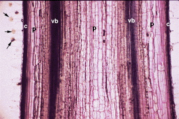
 longitudinal section through petiole (vb-vascular bundle, c-collenchyma, p-parenchyma, arrows indicate trichomes) |
| This is a longitudinal section of the leaf petiole. Note how the vascular bundles are visible on both sides of the petiole. This is because they are distributed in a semi cylinder. We are seeing one plane of that cylinder. |
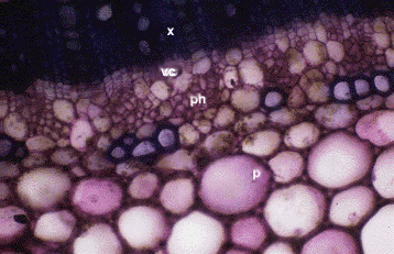
 cross section through petiole (x-secondary xylem, ph-secondary phloem, vc-vascular cambium, p-parenchyma) |
| This petiole is undergoing secondary growth. Notice that the xylem is no longer in distinct bundles but a continuous semi cylindar. |
Introduction | Flowers&Fruit | Roots | Stems | Leaves
Section of Plant Biology Division of Biological Sciences
UNIVERSITY OF CALIFORNIA, DAVIS