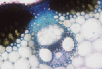
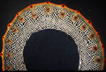


The
internal anatomy of the stem of the rice plant reveals an
amazing world full
of beautiful design, intricate parts, and complex
systems. With
the aid of chemical stains, powerful microscopes, and
polarizing filters,
the following pictures are designed to demonstrate
not only the anatomical
and functional parts of the stem, but also the
beauty in the microscopic
world of the rice plant.
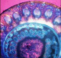
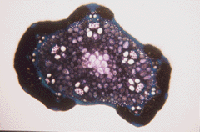
This stem page
will also show the variations
in cross-sections of
the stem, from
the internodal region to below the node to the node
and then through
the panicle.
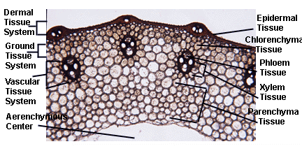
Below is a magnified cross-section
of the outermost region of the stem with the tissues and cells labeled.
Only the cuticle and the epidermal
tissue comprise the DERMAL TISSUE SYSTEM. The other regions are labeled
for a better understanding of the outer components of the stem.
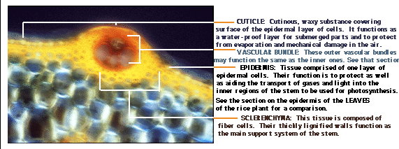
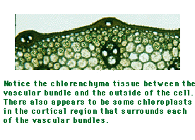
Below are pictures depicting the different regions of the ground tissue system, including the 1)chlorenchyma tissue, the 2) storage parenchyma cells and their contents, and the 3) aerenchymous center of the stem.
1) CHLORENCHYMA TISSUE:
Storage parenchyma cells specified for containing chloroplasts.
The chloroplasts utilize sunlight as energy for metabolizing carbon dioxide
and water for the production of sugars (oxygen as a waste product). The
chloroplasts are what give the stem its green appearance. Notice in the
picture below, that the cortical region of the stem (from the vascular
bundles up to the fibrous layer) does not always contain just chlorenchyma
tissue. The chlorenchyma tissue is primarily toward the outermost portion
of the cortex.
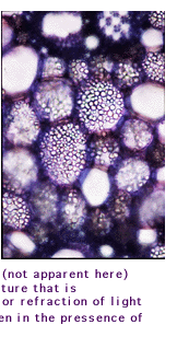
2) STORAGE PARENCHYMA
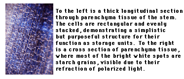
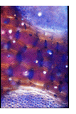
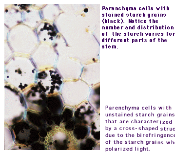
TISSUE: These parenchyma cells specialize is storing contents as
energy in the form of starch and lipids, as ergastic substances in the
form of crystals, and many other substances. The following pictures are
of parenchyma cells as wells as some detail on starch as the stored substance.
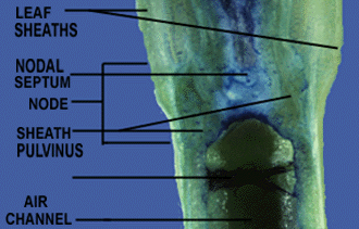
3) AERENCHYMOUS CENTER: The aerenchymous center can be described
as the hollow pith of the stem, although near the nodal regions, the center
becomes filled with aerenchyma tissue. This tissue, although it was not
sectioned for examination, is most likely branched parenchyma cells. The
branch parenchyma cells are also the typical tissue comprising the diaphragm
found in the hollow pith. The aerenchymous center functions primarily as
an air channel for gases to be transported from the aerial plant parts
down to the water-submerged regions of the plant. This demonstrates a
design perfectly fit for the environmental needs of the plant.

The vascular tissue system itself
is comprised of several different tissues with complex functions. There
will be a description and pictures for the 1)phloem tissue and the 2) xylem
tissue in the VASCULAR TISSUE SYSTEM.
1) PHLOEM TISSUE:
The phloem tissue is comprised of phloic fibers, sieve tube members,
and companion cells. The companion cells are extrodinarily distinct in
the phloic tissue. Take a look at the picture below to see the distinct
"big cell, little cell" relationship. The phloic fibers are found primarily
above the phloem, although the outer edge of the rice stem is known to
sometimes have a continuous ring of fiber cells just below the epidermis
consisting of phloic fibers (by definition) as well as "other" fibrous
schlerenchyma tissue.

